|
Monitoring the Neuromuscular Junction This talk evaluates neuromuscular junction monitoring and considers factors affecting nerve stimulation, equipment, the evoked response and its interpretation. Important physiological aspects, not discussed here, include:
ASSESSING NEUROMUSCULAR BLOCK In the anaesthetised patient co-operation is unavailable, hence clinical assessment is limited to surgical comments like, "Can't you make him a little less tight", coughing, straining, patient movement, or changes in lung compliance, all of which only vaguely and non-quantifiably indicate inadequate paralysis. In ocular, micro and neuro-surgery such movement may be hazardous for the patient and excessive doses of muscle relaxants may be given with subsequent inadequate or delayed reversal. Large inter-individual variations in pharmacokinetics and pharmacodynamics of non-depolarising blockers make it hard to guess doses or top-up intervals or use predictive empirical models satisfactorily in these patients.. The clinical use of nerve stimulators has become more widespread (despite their failings) because they permit titration of drug dose to effect, allowing far more accurate prediction of the clinical degree of paralysis than any other means in anaesthetised patients, particularly where deep levels of paralysis are required. Many studies have been performed describing correlations between nerve stimulation responses and the magnitude of clinical paralysis, and the technical and theoretical aspects have been studied in detail, however I am not aware of any published clinical trials documenting outcome improvement from using such devices. Assessing the degree of neuromuscular block depends on the characteristic patterns of response to various muscle relaxants.
CLINICAL ASSESSMENT The following clinical signs are listed in order of increasing block:
Their use is mostly limited to conscious co-operative patients. Sustained headlift, tongue protrusion, and grip indicate that the patient is awake, that tetanic fade is minimal, and that virtually complete neuromusclar junction (NMJ) recovery has occurred. Peak inspiratory pressure correlates fairly well with train-of-four (TO4) ratio. Adequate tidal volume and fairly normal blood gases while breathing spontaneously on an endo-tracheal tube are not sufficient criteria for extubation because at the same time laryngeal and intercostal muscle groups may be very weak. Extubation requires integrity of each component in the respiratory system, so clinical assessment of the anticipated adequacy of ventilation following extubation is essential, and involves consideration many factors beside NMJ function, ie lung function and mechanics, level of consciousness, likelihood of regurgitation, adequacy of local airway reflexes, etc. USING NERVE STIMULATORS 1. The ideal system The ideal system should be cheap, safe, non-invasive, easy to set up, light and portable, allow remote monitoring, and provide information that is reproducible, easily understood, and can be permanently recorded if required. Most importantly, the information should always correlate with clinical findings, allowing accurate prediction of patient response. That no such system exists is not surprising, and explains why so few anaesthetists use nerve stimulators! 2. Nerve Stimulation We all know that the inside of a motor nerve fibre is at an electrical potential of some 70 millivolts below the outside of the nerve, and that if this difference reaches a threshold value, an action potential will be generated which can travel along the nerve and ultimately cause contraction of the muscle fibre it supplies. Some of the factors involved in externally stimulating a nerve include: a) Current Density To cause current to flow through tissue a voltage source must be applied between two electrodes. The total current is proportional to the magnitude of the applied voltage (up to 300 Volts in most commercial nerve stimulators) and inversely proportional to the total skin and tissue resistance (typically 1,000Ω to 10,000Ω), following Ohm's Law (I=V/R). Skin resistance is usually much higher than that of deeper structures and hence it is the greatest impediment to current flow. Furthermore skin resistance is variable from patient to patient, depends on how long the ECG dots have been in place, and whether or not the skin has been properly prepared (by scraping and removing greasiness). Many nerve stimulators are voltage generators and the delivered current is consequently unpredictable from the position of the output knob, so some have digital displays of delivered current. Other units are "constant current" sources (with or without digital displays); in these output voltage changes automatically in proportion to resistance so that delivered current is fixed. The latter are preferable because no user intervention is required if skin resistance changes. All the current that leaves one electrode has to return to the other, and to do so it must pass through either the skin itself, subcutaneous fat, nerves, and nearby muscle and other tissues. Skin and fat have low conductivity (resistance per unit area) and very little current ever passes through them (unless there is a conductive gel "bridge" between the dots. The proportion of total current going through the nerve depends on its resistance relative to the resistance of all the other possible return pathways to the other electrode. Muscle is a particularly good conductor and if it lies between skin and nerve almost all the current "short-circuits" through it, leaving very little to travel through the nerve. Maximal current density is found immediately below each electrode, hence electrodes should be placed as close as possible to the nerve at it's most superficial location and preferably where it is predominantly surrounded by fat. When current flows into the nerve it traverses nerve fibre membranes; each has resistance, and a voltage is thus generated across the membrane proportional to local current density. If the induced voltage is sufficient, some or all of the fibres may depolarise. b) Fibre Size Small (C or Aδ) fibres have electrical stimulation thresholds about 2-3 times that of larger "touch and proprioception" fibres, for example painless senation in transcutaneous electrcial nerve stimulation (TENS). c) Threshold Higher thresholds mean higher current requirements. d) Pulse width Where single pulses are used for nerve stimulation, the amount of current required depends on the pulse width, so that short pulses are less efficient than longer pulses. The relevant factor is current times time. Muscle fibres are about a four times less sensitive than nerve fibres, so direct muscle stimulation can quite easily occur, resulting in problems with the interpretation of the response. With pulse widths of 200μsec direct muscle stimulation is rare under 80 mA. e) Frequency Normal nerves do not respond to very high frequencies or extremely short pulses at all. Tetanic fade is normal at more than about 300 Hz. 3. The evoked response The force generated by stimulated nerve/muscle groups depends on: a) Muscle Bulk. and Contractility (Calcium, resting load, temperature, etc). b) Type of neuromuscular junction , ie facial, diaphragmatic, peripheral. The diaphragm is much more resistant to neuromusclar blocking drugs (NMBs) than peripheral musculature. This means that a healthy person can maintain good gas exchange while sedated and intubated even with only 3 twitches on TO4 testing. If extubated, however, the patient would almost certainly require re-intubation because the laryngeal and ocular muscles are more sensitive to NMBs than peripheral muscles and would be almost completely paralysed. This is a common mistake. If there is any discernable fade in the ulnar nerve then you can be sure that there will be laryngeal and ocular weakness; this may well be present even if you think all 4 twitches were equal. Conversely, the absence of any response to TO4 testing does not preclude a cough because some diaphragmatic function will be retained after T1 is lost. The onset and recovery of block is faster in 'central' muscle groups such as the larynx, jaw and diaphragm, hence full recovery peripherally should mean complete central recovery as well. "Precondtitioning" of the NMJ by recent tetany will obviously modify responses to twitch testing for several minutes. If the patient is "light" and generating their own tetanic stimulation, then what you see on testing is post-tetanic responses and you will tend to over-paralyse the patient. Conversely, if the patient is well anaesthetised and then wakes up, their struggling will cause post-tetanic potentiation and unexpectedly strong movements will occur. For these reasons I personally feel that it is best to use tetanic testing when possible so that this variability is eliminated. c) Adequate nerve fibre activation. Electrode placement is important. The negative or black electrode is much more effective than the positive electrode for some strange reason and should always be placed as close as possible to the nerve at the point where it is most superficial. The positive electrode is indifferent or neutral and can be large in size. By keeping both electrodes as close together as possible and in the line of the nerve muscle artefact is minimised and stimulation efficiency maximal. If anatomical landmarks are poor, it may be useful to use the nerve stimulator to find the optimal point for placement of the negative electrode. This is easily achieved by first sticking the positive electrode in its usual location, and moving the negative electrode over the surface of the skin while stimulating at about 5mA until maximal response is noted. Conventional placement for the ulnar nerve is negative at the palmar aspect of the wrist, proximal to the creases, and lateral to flexor carpi ulnaris, with positive some 6 or 7 cm proximally. The muscle to test for response is the Adductor Pollicis, because it cannot be directly stimulated when electrodes are placed on the forearm. Lateral Peroneal (indifferent on the knee), posterior tibial, and other peripheral nerves may be used successfully, but if the facial nerve is used it generally demonstrates less twitch depression than peripheral nerves and tends to underestimate block considerably. Also it may be difficult to distinguish between neuromuscular responses and direct muscle stimulation artefact. Up to 70 mA per pulse (through the skin) may be required to be sure of supramaximal stimulation, particularly in the obese patient. Skin resistance causes the greatest impediment to current flow so good electrode contact is very important. ECG dots are commonly used, although skin scraping is usually required to get skin resistance below 5 kΩ, and most battery operated devices are lucky to put out more than 60mA into this kind of load. Supramaximal currents should be used so that the small changes in current that occur with changes in skin resistance, battery condition, and load on the circuitry don't affect the number of fibres stimulated and hence the force of contraction. Current is considered supramaximal when further increases show no increase in force of contraction. Ideally the device should deliver constant current at a given setting and have a linear output characteristic, or allow you to measure the delivered current to check this. If the skin resistance is high, in most devices current fails to increase much with turning the output control above a certain point, and this may lead you to falsely believe that the current flow was supramaximal. This applies even to the newer high-output constant current devices. Subcutaneous needles may be used if supramaximal stimulation is not achieved percutaneously. Devices capable of delivering high currents through the body are capable of macroshock if myocardial current densities are great enough. A draft standard has proposed a warning on any device capable of delivering a pulse of 25μC (μC=micro Coulomb; 25 μC = 50 mA for 0.5 msec) across the thorax. Most nerve stimulators can be used as trans-oesophageal pacemakers. Some newer A-V sequential pacemakers may interpret nerve stimulator spikes as a tachycardia and start overdrive pacing ! d) Proportion of functioning end-plates, which depends on:
d) Temperature - hypothermia in the periphery causes:
Note that the presence of NMB's are only one of many factors involved! 4. Assessment of the response a) Observing or palpating twitches This is the simplest method of assessing response and requires a minimum of extra equipment (eliminating recording errors). Assessment of single twitches (ie, 0.1 Hz) require a properly set up force transducer to document control values. Other stimulation patterns use themselves as control and can be assessed by palpation or observation with reasonable accuracy, however it is clear that this is the source of a lot of error in assessing block. b) Mechanical Force Transducers Disadvantages of cost, complexity, and setup time limit their use. A simple dedicated force transducer is available from Professional Instruments (sold by Anaesthetic Supplies P/L) and this simplifies things considerably. Attention to detail is necessary to minimise errors, but accurate assessment of muscle responses, paper printout, and remote monitoring are advantages, particularly in research. Tetanic stimulation of the ulnar nerve can cause 5-6 Kg force in the adductor pollicis brevis, so the transducer must be capable of measuring up to 10 Kg (ie, Grass FT10) if you want to look at tetanic responses. Secure fixation to the arm and correct orientation of the transducer are necessary to prevent baseline drift and nonlinear response. Preload should be adjustable to around 200g. c) Accelerometers Accelerometers are small electronic devices which transduce acceleration into electrical potentials and can be easily sticky taped to the thumb. F=mA so for a given mass of thumb the acceleration is proportional to applied force. Output correlates well with force transducers (although they cannot respond appropriately to tetany) and there is no need for pre-tensioning and calibration is simple. A device similar to the Myotest is available using this system. c) Integrated EMG New devices capable of both stimulating and recording the integrated EMG are becoming available, and as they only require placement of 5 ECG dots (2 stimulating, 2 recording, 1 neutral) are simpler to set up. With non-depolarising relaxants the EMG underestimates the block both in twitch amplitude and TO4 ratio, so that it is more useful in asssessing depth of block, rather than reversibility. The opposite is true of suxamethonium. EMG devices avoid problems with transducers but are susceptible to diathermy and have considerable problems if skin resistance changes occur. Both these problems will probably be overcome in the near future and I think they will become more widely used. Be aware of the tendency to underestimate nondepolarising block with the EMG.
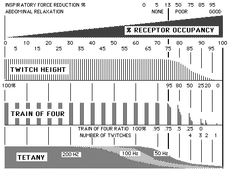
Receptor occupancy and clinical responses 5. Stimulation patterns and their uses a) 0.1 Hz twitches Single twitches repeated every 10 seconds require control values for valid comparisons, may appear normal with considerable weakness, are a poor indicator of deep paralysis, and require the use of force transducers for meaningful data. Most research work on dose-response studies uses this pattern. b) Train of four (TO4) Fade is prominent with non-depolarising blockers and at 0.5 Hz is greatest by the 6th twitch. Using four twitches at 0.5 second intervals (TO4) was popularised by Ali and from these the ratio of T4/T1 (the "TO4 Ratio") can be derived. The degree of paralysis is estimated from the number of twitches present, or if four are present the TO4 ratio. The obvious advantage is that responses can be easily quantified without the use of force transducers as the first twitch is used as a control for the others. Counting the number of palpable twitches is quite a good guide to deeper levels of paralysis; two or more twitches usually implies reasonably easy reversal and some return of muscle tone, while virtually no response suggests difficulty with reversal, weak cough at best, and very little muscle tone. When visual or tactile means are used to assess response, TO4 ratios around 0.25 are commonly estimated at between 0.1 and 0.7, while at 0.5 some 40% of and at 0.7 fewer than 10% of observers can reliably detect any fade at all. Consequently the presence of any detectable fade indicates the presence of some paralysis and furthermore even if all four twitches appear normal many patients are in fact partly paralysed. Hence the train of four is most useful to assess block associated with general surgical levels of paralysis where the aim is to keep only one or two twitches visible for absence of abdominal wall tone, and also to assess whether or not a patient is easy to reverse (usually if four twitches are present there is no problem at all) It cannot be used to assess very deep levels of block (no T1!) and is not very sensitive to assessing adequacy of reversal. c) Dual Burst Stimulation (DBS) Described recently by Viby-Mogensen as a 50Hz train of 3 repeated 0.75 seconds later by an identical train of three. Each group of three twitches results in one twitch, and hence only two twitches available for comparison. Since the first twitch sums T1, T2 and T3, while the second sums T4, T5, and T6, it is easy to see how the presence of fade would be easier to notice and there is data to support this. As the level of block increases, response to the seond burst is lost as the third twich of TO4 is lost; the first burst is retained until a little after you lose all response to TO4. Surgical paralysis is generally OK if only one response is present; the patient is reversible if two are present, particularly if the second is strong. TO4 is better for quantifying the intensity of "surgical" paralysis, whereas DBS is better for noting persistance of fade after reversal. If you use NMB's so that there is just no response to DBS, the patient will be a little more paralysed than if there was just no response to TO4. d) Tetanic stimulation Continuous stimulation at either 50 or 100 Hz is so painful as to preclude its use in conscious patients, and is difficult to quantify, but is probably the most useful and emulates physiological maximal responses. Tetany is more sensitive to both residual and deep paralysis than any other form of monitoring. The presence of any persisting strength during tetany is a good indicator of the patient's ability to maintain muscle tone. Comparing two bursts of tetany (each 3-5 seconds long) with a gap of 3 seconds results in post-tetanic potentiation of the response to the second burst. When assessing adequacy of reversal the initial part of the second response (potentiated) can be compared to the last part of the first (faded). If fade is present it is becomes more obvious with this rather than any other method. e) Post-Tetanic Count (PTC) This consists of counting 1 Hz twitches 3 seconds after 5 seconds of 50Hz tetany and can give an approximate time to return of response to single twitches and hence permits assessment of block too deep for any other technique. A Post-Tetanic Count (PTC) of 2 by palpation suggests no twitch response for about 20-30 minutes, PTC of 5 about 10-15 minutes. This is clearly the best method for monitoring paralysis for patients in whom you seek to prevent diaphragmatic movement, ie micro-neurosurgery; it is best to use infusions of drugs and aim for PTC of 2. f) Clinical situations General-purpose monitoring: TO4 is perhaps best. NB: reversal drugs are most effective if the ratio of antagonist to antagonist is most favourable. If your patient seems quite paralysed and you want to reverse them, it's best to wait for at least 25% return of twitch height (say 2-3 twitches) for the fastest return of strength; given any sooner the level of reversal will be too low at the end. UNUSUAL CLINICAL SITUATIONS a) Hemiplegia - resistance to non-depolarisers within 2-3 days on affected side, possibly due to loss of cerebral inhibition. Always monitor hemiplegic patients on the unaffected side. b) Parkinsons Disease, Multiple Sclerosis, Tetanus, Intracranial Lesions - normal sensitivity to non-depolarisers. c) Paraplegia and Quadriplegia - increased sensitivity to non-depolarisers. The difference in response of the NMJ for upper and lower lesions suggest that extrajunctional chemosensitivity is not involved. (It is responsible for the hyperkalaemia following suxamethonium). May also happen following burns, immobility, prolonged administration of NMB's, etc. d) Amyotrophic Lateral Sclerosis, Polio - increased sensitivity to non-depolarisers. e) Peripheral Neuropathies - usually normal response, although patients with neurofibromatosis may be sensitive. f) Myotonias - usually normal response to non-depolarisers, occasional sensitive patients. g) Muscular Dystrophies - mostly normal responses except in the "Ocular" type, which is very sensitive to non-depolarisers. Duchenne may be a risk factor for MH.
BIBLIOGRAPHY 1. Ali HA, Savarese JJ: Monitoring of Neuromuscular Function. Anesthesiology, 45: 216-246,1976. 2. Bowman et al. Prejunctional and Postjunctional Cholinoceptors at the Neuromuscular Junction. BJA, 82: 1111,1980. 3. Ali HA et al: Twitch, Tetanus, and Train-of-four as Indices of Recovery. Anesthesiology,54: 294,1981. 4. Kopman AF, Lawson D: Milliamperage Requirements for Supramaximal Stimulation of the Ulnar Nerve with Surface Electrodes. Anesthesiology 61: 83-85, 1984. 5. Howard-Hansen P et al: Tactile Evaluation of the Posttetanic Count. Anesthesiology, 60: 372-374, 1984. 6. Viby-Mogensen J et al: Tactile and Visual Evaluation of the Response to Train-of-four Nerve Stimulation. Anesthesiology, 63: 440-443, 1985. 7. Mylrea, KC: Evaluation of Peripheral Nerve Stimulators and Relationship to Possible Errors in Assessing Neuromuscular Blockade. Anesthesiology, 60: 464-466, 1984. 8. Jones, RM: Neuromuscular Transmission and its Blockade. Anaesthesia, 40: 964-976, 1985. 9. Viby-Mogensen J et al: Post-tetanic Count (PTC): A new method of evaluating intense non-depolarising neuromuscular block. Anesthesiology, 55:458, 1981. 10. Graham GG, Torda TAG et al: Relationship of TO4 Ratio to Twitch Depression during Pancuronium Induced Neuromuscular Blockade. Anesthesiology 65: 579-583 1986
Last updated Tuesday, December 15, 2020 |
|||||||||||||||||