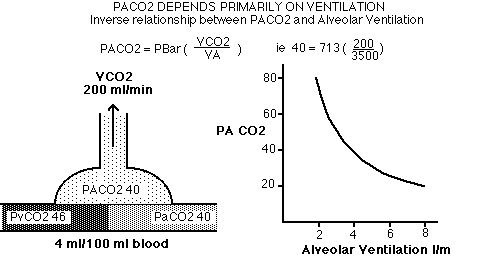
Chris Thompson USyd Lectures Listing VENTILATION, VENTILATORS and HUMIDFICATION INTRODUCTION Normally, Alveolar Ventilation is unconsciously regulated to maintain constant arterial blood gas tensions (particularly CO2), despite variable levels of oxygen consumption and CO2 production. Many drugs and techniques used in anaesthesia interfere with control or mechanics of ventilation, and it is the Anaesthetists responsibility to ensure the adequacy of ventilation during the perioperative period. Ventilation and ventilators are consequently of great importance to the Anaesthetist. Correct use requires a good understanding of basic respiratory physiology as well as how the individual ventilator operates. I've recently uploaded a plain text abstract and PDF set of slides from a recent talk on ventilators, optimising PEEP and lung recruitment. Basic Principles Venous blood always has a lower PaO2 (40 mmHg or 75% saturated or 15 ml O2/100ml blood) and higher PaCO2 (46 mmHg) than inspired gas (PiO2 150 mmHg, PiCO2 usually 0). Hence there is a partial pressure gradient driving Oxygen in and CO2 out of the pulmonary capillary blood. Ventilation of the lungs is the pprocess that mixes fresh inspired gas with alveolar gas. If there is no ventilation at all, there will be no replenishment of oxygen and no removal of CO2. PAO2 will fall and PACO2 will rise towards the venous O2 and CO2 tensions. After the onset of apnoea, CO2 rises rapidly and within about 30s it is about 50mmHg. This causes nearly all of the air hunger experienced while holding your breath. The subsequent rate of rise of CO2 tension is much slower, thaking 15 minutes or so to reach 80mmHg, because it is very soluble. The rapid response of arterial CO2 to apnoea explains why it is such a good gas for our body to use as the primary control mechanism for ventilation. In contrast a reservoir of oxygen exists within the alveoli that can maintain acceptable oxygen tensions for about a minute. If the lung volume is adequate, after a deep breath, and in particular if the lung is filled with a higher than usual oxygen partial pressure, acceptable alveolar oxygen tensions can be maintaned for much longer. However, should FRC be reduced, oxygen consumption increased, or collapse likely, the benefit of 'pre-oxygenation' is markedly reduced. If the ventilation is greater than is needed, then the alveolar gas tensions will be shift closer to inspired gas, ie the CO2 level will be lower, and the oxygen level a little higher. Non ventilated but perfused (very low V/Q) lung units shunt venous blood to the arterial circulation, causing hypoxaemia that cannot be corrected by increasing FiO2. The matching of ventilation to perfusion across the majority of alveoli, even in the presence of lung disease, is the key to optimising ventialtion. Definitions Ventilation is the process by which Oxygen and CO2 are transported to and from the lungs. Tidal Volume (Vt) is the amount of gas expired per breath - typically 500ml at rest. Deadspace Volume (VD) is the sum of the Anatomic Deadspace, due to the volume of the airways (typically 150ml), and Physiologic Deadspace, due to alveoli which are ventilated but not perfused (usually insignificant). Minute Volume (VE) is the amount of gas expired per minute. Alveolar Ventilation (VA) is the amount of gas which reaches functional respiratory units (ie, alveoli) per minute. VA = (Tidal Volume - Deadspace) x Respiratory rate Lung Volumes
Lung Mechanics Inspiration An active process requiring musular effort; 75% diaphragmatic at rest; intercostals used on exertion. Inspiratory effort causes:
Maximum inspiratory force sometimes used as an index of resp. effort; if < 20 cmH2O most patients have difficulty Expiration Usually a passive process due to lung recoil:
Respiratory Rate and I:E ratio Normal respiratory rate is about 15 breaths per minute, increasing markedly with exertion. Normal I:E ratio at rest and while asleep is 1:2 or less. On exertion the I:E ratio is 1:1. Inspiration is normally an active process (requiring work). Expiration is passive, and usually longer than the time required for exhalation, resulting an a no-flow period. When breathing spontaneously, the work of breathing is minimised by keeping inspiratory times short and tidal volumes low - just enough to get rid of the produced CO2. To minimise collapse, sighs are taken from time to time. Under anaesthesia, FRC is reduced, resistance increased and collapse is common. Longer than normal inspiratory times may be required forall alveoli to reach inspiratory equilibrium. Expiratory pauses with no gas flow will not contribute to ventilation (no gas is flowing) and may contribute to collapse. All work of respiration is done by the machine. It is likely that the optimal I:E ratio under anaesthesia is 1:1, not 1:2. A good starting point is a respiratory rate about 12 at an I:E ratio of 1:1. The actual inspiratory and expiratory time should be adjusted so that the gas flow curve in pressure mode shows near complete equilibration both in inspiration and expiration. Airway Resistance
Intrapleural Pressure
Compliance "Static" Compliance is a measure of the "stiffness" of lung and chest wall, typically 50 ml/cmH2O in adults and proportionally less in kids. It is usually due equally to lung and chest wall compliances (100 ml/cmH2O each). Surfactant improves lung compliance, especially at low lung volumes; its absence as in ARDS, results in stiff lungs and a tendency for the alveoli to collapse and fill with fliud. Lung collapse results in a reduction in available ventilatable lung volume, reducing compliance and causing higher than expected airway pressures. "Dynamic" compliance includes the extra pressure needed to overcome resistance to airflow, inertia of chest wall, and viscoelasticity of tissues. Total compliance varies from person to person and from time to time. A ventilator with pressure limited inspiration will deliver varying tidal volumes during an anaesthetic and from patient to patient. Most modern anaesthesia ventilators are of the "Volume Preset" type to minimise this problem. Work of Breathing Work = Pressure x Volume Respiratory work at rest or during exercise is seldom responsible for more than 5% of the total body work. Most of this is used to overcome the lung and chest wall stiffness during inspiration. Work to overcome airway resistance is usually very small, except during exercise or in athsmatics. Patients with most respiratory diseases have increased respiratory workloads, which may be due to high respiratory rates, stiff lungs, or high airway resistances. When the patient becomes so exhausted that they can no longer keep up the workload, respiratory failure ensues. Anaesthetic machine tubing, one-way valves, and ETTs all increase total resistance and respiratory work, while drugs will diminish respiratory effort, so that the patient with poor respiratory function usually requires ventilating both during and after the operation. CO2 Elimination Oxygen Transport
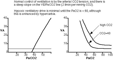
Effect of Shunts Some venous blood passes through the lungs without equilibration with Alveolar gas. This "Venous Admixture" or "Shunt" subsequently mixes with oxygenated blood in the pulmonary veins, and has the effect of reducing PaO2 and elevating PaCO2. While the slight rise in PaCO2 can be overcome easily by increasing the ventilation to normal alveoli, the same is not true for PaO2. Normal alveoli can blow off twice as much CO2 as usual if ventilated twice as much normal, but never saturate the blood leaving them any more than 100%. A pure shunt causes hypoxaemia that DOES NOT correct by increasing inspired oxygen. A patient with a 50% shunt breathing 100% inspired oxygen will only get a PaO2 of about 60 mmHg, but doubling their ventilation will maintain normocarbia. Low V/Q ratio Areas of lung with lower-than-normal ventilation cause hypoxaemia. The blood leaving these areas is part-way between alveolar and mixed venous. If we reduce the total ventilation of an otherwise normal lung by half, ie give it a global V/Q ratio of 0.5, CO2 levels will rise and eventually reach equilibrium at 80mmHg but the patient will become very hypoxic well before the CO2 levels get high. Increasing inspired oxygen to as little as 30% will, however, completely correct the resulting hypoxaemia. Thus hypoxaemia due to hypoventilation can be easily corrected with supplemental oxygen, whereas that due to true shunt will not correct no matter how much oxygen is administered. Control of Ventilation
Effects of Anaesthesia
Collapse, atelectasis and low V/Q areas are common under anaesthesia. As little as 30-40 seconds of apnoea in an oxygen rich environment can cause significant collapse. Recruitment manouvres can expand collapsed lungs, but PEEP alone cannot. However, after recruitment, optimal PEEP can maintain an 'open lung' and prevent or minimise subsequent collapse. Recruitment manouvres under anaesthesia can cause hypotension, especially in hypovolaemic or elderly patients, and will increase venous pressure, so they should be performed at an appropriate moment and perhaps with transient vasopressure support.. Typical approaches are to manually valsalva for 30s to 30-40 cmH2O pressure, or to keep ventilating as usual but add say 20-25 of PEEP for about 30s with a maximul pressure limitation of about 45. The time component is essential. Frequently compliance will improve by about 1/3 after recruitment. Hypoxia in the recovery room or PACU is much less of a problem if the patient's lungs are recruited before the end of surgery, if the patient is extubated sitting up, and if periods of apnoea in high oxygen environments are avoided. This is of particular importance with obese patients Classification
Effects of Artifical Ventilation Respiratory:
Cardiovascular:
Renal
Pressure Support, CPAP or BiPAP PEEP alone is not good for spontaneous breathing, because the patient cannot breathe 'in' easily in the expiratory phase. Simple PEEP generators are just an APL type valve occluding the expiratory limb, with the bag as a poor pressure reservoir. These simple systems will maintain FRC during controlled ventilation but markedly increase the work of breathing during spontaneous respiration. CPAP means 'constant positive airway pressure', typically meaning that the ventilator can maintain constant airway pressure during the expiratory phase, so that the patient can breathe in at any time during the expiratory phase without the airway pressure falling below the CPAP level. Patients that require PEEP while breathing spontaneously require a CPAP generator. BiPAP refers to maintaining constant airway pressure during both inspiration and expiration. This is a variant of Pressure Control ventilation in which the patient can breathe in and out during any phase of respiration. Common BiPAP modes have the ventilator synchronise both inspiration and expiration to patient effort. Inspiration usually commences on detection of a patient-generated trigger (typically flow or pressure). The ventilator switches to expiration when the flow rate during inspiration falls below the peak flow rate (tyically below 25% of peak flow). Pressure Support ventilation modes under anaesthesia are forms of BiPAP often with a fall-back in case of apnoea. Dräger Pressure Support machines will dynamically switch in and out of a fixed rate form of BiPAP ('apnoea mode') at a predertmined rate whenever apnoea occurs. The advantages of BiPAP/Pressure Support are
Optimising PEEP or CPAP during controlled ventilation is relatively easy. When a ventilatiator is in Pressure Control Mode the tidal volume resulting from a given differential airway pressure is proportional to lung compiance. Improved tidal volume at the same differential pressure indicate an improvement in lung compliance. If the inspiratory and expiratory times are long enough for alveolar equilibration, optimal PEEP (in a mechanical sense) is that which results in the greatest tidal volume at a given differential airway pressure. After recruitment, PEEP should always be optimised. The ideal setting may change. My approach to recruitment and PEEP optimisation is to:
Ventilators Classification
Use in Anaesthesia
With CO2 absorber ON:
With the CO2 absorber OFF:
Hazards
Monitoring Ventilation
Physics
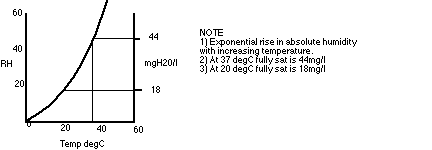
Measurement of Humidity
Physiology The nose is a very efficient humidifier:
Mouth-breathing is less efficient (60% RH in the upper trachea) Heat and water loss through the nose is minimised by cooling on inspiration and warming on expiration. Humidification is required to maintain of ciliary activity, prevent squamous epithelial changes (Mucosal changes in 2-3 hours), prevent dehydration and thickening of secretions and possible ETT obstrucion, minimise atelectasis and tracheitis, and to decrease heatloss Methods ANAESTHETIC CIRCUIT CONSIDERATIONS
HEAT AND MOISTURE EXCHANGERS
HUMIDIFIERS Up to 100% humidification, essential for longterm respiratory care or if drying of excessive secretions occurs despite HME's. Modern types heat both the water bath and patient hose to prevent rainout. Disadvantages:
EQUIPMENT FISHER & PAYKEL
GRANT - NICHOLAS
BOURNS
NEBULISERS
For more detail, see my humidification notes. Last updated Tuesday, December 15, 2020 |
||